Reditus offering new test for bladder cancer
Reditus Laboratories is starting to use a bladder cancer test that is more sensitive and specific than other tests for the disease.
UroVysion is a fluorescence in situ hybridization (FISH) assay developed for the detection of bladder cancer in urine specimens. UroVysion detects urinary cells that have chromosomal abnormalities consistent with bladder cancer.
“It’s used to help monitor patients with a history of bladder cancer and to detect new cases,” said Reditus Laboratories Cytogenetic Technologist Mark Potts said. “There is a high incidence of bladder cancer out there and this is a tool in early detection.”
Bladder cancer is the fourth most common cancer among men but is less common among women, according to the American Cancer Society. The earlier that cases are detected, the earlier that treatment may begin, Potts said.
UroVysion is used in conjunction with urine cytology, Potts said. If urine cytology reveals suspicious cells, UroVysion may be ordered. UroVysion is more specific because it looks at the chromosomes within the cells.
“It looks at specific gene sequences in chromosomes 3, 7, 17 and 9p21,” Potts said. “We are looking for a complete loss of 9p21 and gains in chromosomes 3, 7 and 17.”
UroVysion is a multi-step process. A summary follows.
The process begins, at a clinician’s office, when a patient gives a urine sample. A preservative is added to the sample, which is shipped by courier to Reditus.
At Reditus, as shown in photo 1, the urine is spun down to make a cell button at the bottom and a buffer is put in to suspend the cells, Potts explained. After it’s spun down again, a solution is added to preserve the cell.
Three concentrations of cells are placed on a glass slide to air dry, as shown in photo 2.
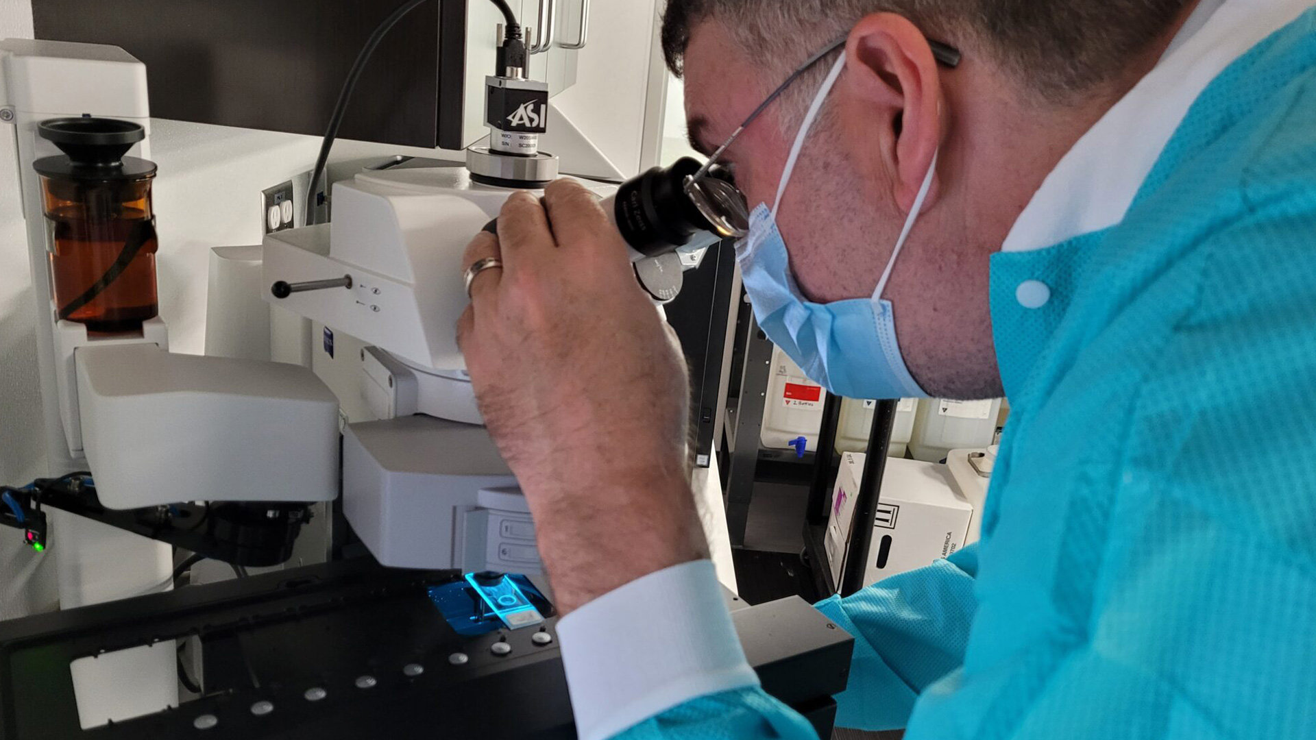
The slide is analyzed under a phased contrast microscope, as shown in photo 3, and the most populated concentration is selected for the FISH procedure.
The slide is immersed in a hot protease solution, as shown in photo 4. “That breaks down the cell membrane so we can get to the nucleus,” Potts said.
The cell is then dehydrated. The actual FISH process then begins as the slides are denatured, meaning the DNA strands are unzipped so probes can get in and find matching sequences within the DNA strand. When exposed to fluorescent light, each chromosome gives out a signal, each of which is a different color, and the color tells whether there are more than two copies of the chromosome in that cell, as shown in photo five.
“If we see gains in 3, 7 and 17, in greater than or equal to four cells in the analysis, then that is likely a positive result for bladder cancer,” Potts said. “Or, if we see a complete loss in 9p21, in greater than or equal to 12 cells in the analysis, that also is considered a positive result.”
After 18 hours, the slides are placed in wash solutions, dried, then a reagent is put on the slides, as shown in photo 6.
The slide is placed in a freezer for 30 minutes, warmed up, then put on a scanner scope, which scans for urothelial cells, as shown in photo 7.
“When it’s done, the technologist scores the cells and determines whether they’re normal or abnormal,” Potts said. “A print report goes to the pathologist to be signed out and reported to the physician.”