Reditus Histology Technicians Serving Podiatrists, Expanding Into Urology
By Paul Swiech

Reditus Laboratories’ histology technicians help doctors to diagnose and treat diseases by examining tissue at the cellular level.
Histology is the study of tissues under a microscope. Through histology, a pathologist can detect cell or tissue abnormalities that may signify cancer, disease status, or detect microorganisms that may be responsible for infection.
The Reditus histology lab, which received its first live specimens in November 2019, serves mostly podiatrists from seven states. Specimens that histology technicians examine include toenail clippings and skin biopsies to look for fungal infections, wound samples, dermatitis and wart specimens.
“If it’s a wart or pigmented lesion, we’re trying to rule out cancer or melanoma,” said Kela Rodeffer, Reditus histology general supervisor. “For podiatrists (overall), we’re helping to diagnose everything from ingrown toenails to cancer.”
The Reditus histology lab is expanding into urology.
“We recently started receiving urology specimens, so far, just from doctors in Illinois,” Rodeffer said. Urology specimens include bladder biopsies and prostate biopsies to rule out cancer, she said.
“We will soon accept urine to test for bladder cancers,” she said.
“We are going to start performing Fluorescent In-Situ Hybridization (FISH) analysis soon, which is very exciting and completely new to central Illinois,” Rodeffer said. “It is a highly sensitive method for bladder cancer detection that we will offer our urology clients. We can take a urine specimen and label any cancer cells present in it with a fluorescent probe that makes them visible by microscopy for a pathologist. It’s very robust technology and we may use it for other types of testing in the future. We hope to have that going by mid-summer.”
The process in the histology lab begins when technicians receive a specimen and enter patient diagnostic, demographic and insurance information into the Reditus laboratory system. This information is required by regulatory agencies. The specimen is given an accession number, which is used to identify the specimen throughout the process.
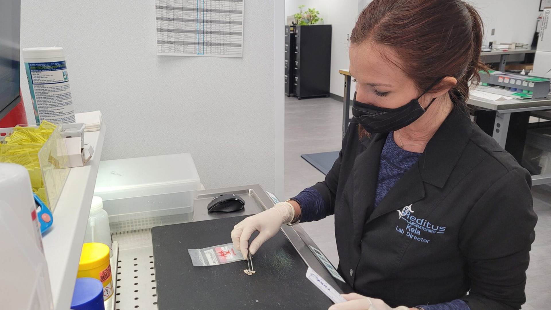
The specimen proceeds to a grossing station, where technicians, as shown by Rodeffer in the accompanying photo, examine the tissue, take measurements and describe what they observe, noting the size, color, texture and abnormal features of the tissue.
“The observations we make during the gross examination are recorded in our computer system and are available electronically to the pathologist during his review of the slides,” Rodeffer said.
A sample of specimen is transferred to a cassette which goes into Formalin solution for preservation. Toenail specimens go through an additional step of de-keratinizing to soften the sample prior to processing. Then, a rack of specimens in a cassette goes into a tissue processor, which chemically processes the tissues and infiltrates them with paraffin wax, as demonstrated in the photo by Rodeffer.
The next step in the process is the embedding center, where histology technicians like Hannah Finnegan take tissues and put them in a mold that will result in a wax block embedded with specimen tissue.
After the wax cools, what results is a solid block of paraffin with tissue embedded inside. A sample is held in the accompanying photograph. This makes it possible to cut the tissue sample.
Next, Finnegan uses a microtome to cut ribbons of sections of the block that are four microns thick, or the thickness of one cell.
The tissue is placed in a water bath to smooth out the tissue. Then Finnegan separates each section of tissue and transfers the sections from the water bath to a glass slide.
Next, slides are placed in an oven for one hour for heat fixation to make sure the cells adhere to the slide. Then the slides are stained, as illustrated in the photograph, to show the different components of the cell. Different stains are used to identify fungal and bacterial infections or cancer.
Finished slides are placed in a tray, as shown in the accompanying photograph. A final quality control check is performed and the slide is matched with the original paperwork and delivered to the pathologist for review and diagnosis.
Newer to Reditus Laboratories is immunohistochemical (IHC) staining, which is a more precise stain that can detect specific cell antigens. Preparing IHC slides in the accompanying photo is histology technician Lorin Heiser.
“It’s a highly specific, highly sensitive test,” Rodeffer said. “We will be using it as a diagnostic tool for cancers, particularly prostate cancer and melanomas.”
“The whole process, from specimen receipt to finished slides, takes about 24 hours,” Rodeffer said. “The slides go to the pathologist, who usually read the slides and finalize the case fairly quickly, but occasionally there is a need for further chemical stains or immunohistochemical studies to make a proper diagnosis, which adds a bit more time.”
The pathologist reports their findings to the doctor who sent the specimens.
While most hospitals and some private medical practices have histology labs, Reditus Laboratories is the only lab in central Illinois performing specialized work on podiatry and urology specimens.
“The kind of specimens we handle at Reditus are technically challenging and require highly skilled histology technicians,” Rodeffer said. “Histology is as much an art form as it is a science and the Reditus histology technicians are incredible artists. The fact that we are going to bring FISH testing in-house makes us truly unique to the area.”


